
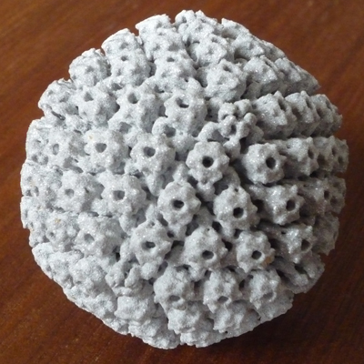
Shapeways community member David
Bhella 3D printed a 5 CM model of the Herpes Simplex Virus. The
Herpes was as a gift for a retiring professor. Presumably, said
professor is one of only a small group of people that are glad to have been given herpes. Intrigued I
asked David to tell us more about what he does at the MRC Virology Unit,
Institute of Virology, University of Glasgow with 3D printing.
Joris Peels: Why are you interested in 3D printing?
David Bhella: I am interested in all aspects of 3D technology (rapid
prototyping, 3D
displays and commercial 3D animation software), as my work is entirely focused on solving the structures of viruses in three-dimensions and I
find visualisation in 2D media deeply unsatisfactory.
I think that rapid prototyping is a really interesting way of allowing
one to appreciate the complexity and symmetry of viruses. Holding the
model in your hand is such a ‘human’ way of understanding an object.
Unfortunately the potential for understanding these structures at
high-resolution cannot be realised in this manner, because proteins are
extremely complex molecules, and a virus is a complex assembly of
proteins. So, the Herpes model I have printed through Shapeways is
comparatively low-resolution (about 2.5 nanometers resolution – the
object itself is 125 nanometers in diameter). We are now working at
better than one nanometer resolution. At this level the 3D shape of
individual protein molecules becomes visible, showing us how they fold
up. To show this as a polygon surface becomes less meaningful then, and
we have to start looking at more complex means of visualisation, also
the poly count becomes so high that commercial 3D software cannot handle
it.
For the moment then, the strength of 3D printing is in teaching and in
public engagement (and in bespoke gifts for retiring professors). I am
really enthusiastic about the prospect of producing large metal
sculptures of virus particles that people can handle and walk around, I
think that the beauty and symmetry of viruses really highlights the
elegance of nature and evolutionary processes. As I am the scientist
responsible for public engagement in my department, I am lucky enough to
be able to dedicate some of my time to this area. Furthermore, my wife
is head of the science team in our local science museum (Glasgow Science
Centre), so I have access to a great venue for P.E activities, which is
staffed by motivated and enthusiastic science communicators who can
help me. A couple of years ago we created an art exhibition called
molecular machines (http://www.molecularmachines.
got a lot of media attention. I think it would be great to produce a
molecular machines 2.0 that exploited the latest in 3D panel displays
and 3D printing.
Joris Peels: What do you do?
David Bhella: I work on many aspects of the virus life-cycle using
cutting-edge
electron microscopy and image processing techniques to understand the
structures of viruses in three-dimensions. Viruses are the smallest of
pathogens to infect man and range from ~30 – 500 nm in size (A nanometer
is 1/1000th of a micrometer which is 1/1000th of a millimeter). They
reproduce themselves by invading our cells and hijacking the cellular
machinery to make thousands more viruses particles. As they have a very
small number of genes, they assemble from multiple copies of only a few
types of protein. They are therefore highly symmetrical, employing
either helical or icosahedral symmetry to make a shell (called a capsid)
that ferries their genome between cells while protecting it from
damage.
So, we take 2D images in the transmission electron microscope and use
image processing methods to average together images of thousands of
particles into a 3D reconstruction. I attach a raw image from the
microscope of a
Feline calicivirus. I am interested in this virus as it is from the same
family of viruses as the noroviruses that cause winter vomiting
disease.
Joris Peels: Was it just fun?
David Bhella: It is always fun!
![]()
Why am I reminded of scenes from the movie “Attack of the Killer Tomatoes”??
Kind of creepy, but at least you are not printing in ashes!
When I was in high school, one of my fellow students gave herpes to the vice principal on a school camp.
He did not ‘retire’ but did take some time off after that.
A firend of mine who is a nursing student caught the common cold (plushie virus) at a carnival event on campus! :D.
After trying several sites myself, I just wanted to say that anything is possible, like finding the love of your life on Hsoulmate.com even you have herpes,HPVor HIV.