


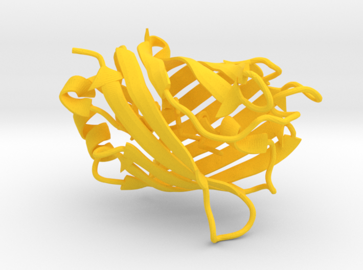
Citrine Fluorescent Protein
Made by
Have a question about this product?
contact the designerProduct Description
Say hello to some of the brightest things under the microscope!
Have you ever seen an animal glow? Maybe you live in a place where you can spot fireflies in the summer, flickering on and off in a field. Maybe you're lucky enough to live near a beach that ocassionally glows blue from plankton or other sea beasties. When living creatures emit light we call this bioluminescence. Different species use different strategies to glow, but easily the most important light-up animal for science is the jellyfish Aequorea victoria. This jellyfish lives in the Pacific ocean and has a thin frill that glows green in the dark from Green Fluorescent Protein, or GFP. Today fluorescent proteins are an essential part of any cell biology lab, but it took an incredible effort from a collection of very bright minds to turn this unassuming jellyfish molecule into a powerful tool for science. In fact, this effort was rewarded in 2008 by the Nobel Prize in Chemistry, given to three key people who drove the research on fluorescent proteins over more than 50 years to where it is today. Each contributed to the story in a unique way, and I recommend the Nobel Prize speeches by these scientists to everyone who is curious [1,2,3].
The man responsible for discovering GFP and its unique properties is Dr. Osamu Shimomura, a Japanese scientist who studied a crustacean that uses a two-part system to glow blue; a protein called luciferase and its small molecule partner, a chromophore called luciferin. In the presence of oxygen, the luciferase protein chemically alters its partner molecule, releasing blue light as part of the reaction. Fireflies also use a version of this system to flash yellow [4]. Shimomura moved to the United States in 1960 to solve the puzzle of what causes Aequorea to glow green. He traveled to Washington with a team of researchers to collect jellyfish and literally squeeze out their secrets. Hundreds of hours (and thousands of jellyfish) later, he isolated a protein he named aequorin that could be stimulated by calcium to shine... blue?
The green glow of the jellies actually came from a different protein, which took another 16 years to successfully isolate. This Green Fluorescent Protein could absorb the high-energy blue light produced by aequorin and emit a green photon of lower energy [5]. What makes GFP so special is that it could emit light without any small molecule as a helper; luciferase needs luciferin, aequorin needs coelenteramine, but GFP came with its chromophore built-in!
This fact sparked the interest of Dr. Martin Chalfie, who successfully took the DNA sequence for GFP and showed that it could be placed in other organisms and still do its job: blue light in, green light out [6]. His lab studies the development and behavior of a transparent worm called C. elegans. GFP can be copied and pasted into the worm's genome so that it gets expressed in certain cells, like neurons. Shining a blue laser on these worms will make the neurons glow green. GFP can even be added to the ends of other proteins, creating a glowing "tag" that shows exactly where that protein is expressed in a living organism. After publishing his results, Chalfie was flooded with requests from scientists interested in using the gene. But Dr. Roger Tsien took this bright idea to a whole new level, breaking down the physics and chemistry of fluorescent proteins to engineer a rainbow of glowing molecules for biologists to use.
Tsien was entranced by glowing molecules and spent years studying the chemistry of electrons jumping from one biological molecule to another in order to make a tool for studying cells. After learning about GFP, he wished that the protein came in multiple colors so that he could track multiple proteins at the same time. His lab mutated GFP to produce a variety of greens and blues that could tolerate different conditions of salt, pH and temperature [7]. A few years later it was discovered that corals use a modified version of GFP as a pigment that glows red. Unfortunately this pigment wasn't useful as-is; in coral, four copies of the Red Fluorescent Protein stick together as a tetramer, and their stickiness ruins any chance of using it to tag proteins in living organisms. So the Tsien lab mastered the art of directed evolution, causing random mutations in the gene and then selecting mutants from the pool that have desirable features like losing the tendency to stick together [8]. It wasn't long until a rainbow of fluorescent proteins was evolved for scientists to use, and they have transformed the way we research development, biochemistry, disease, and practically every other facet of biology.
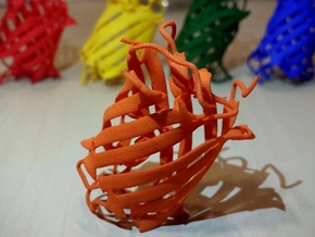
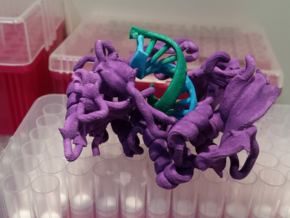
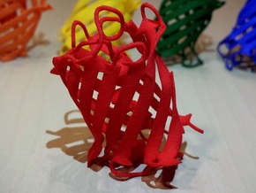
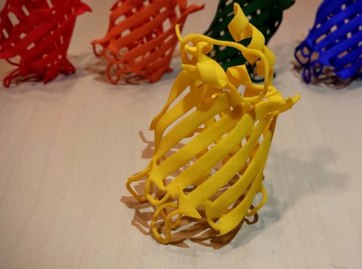
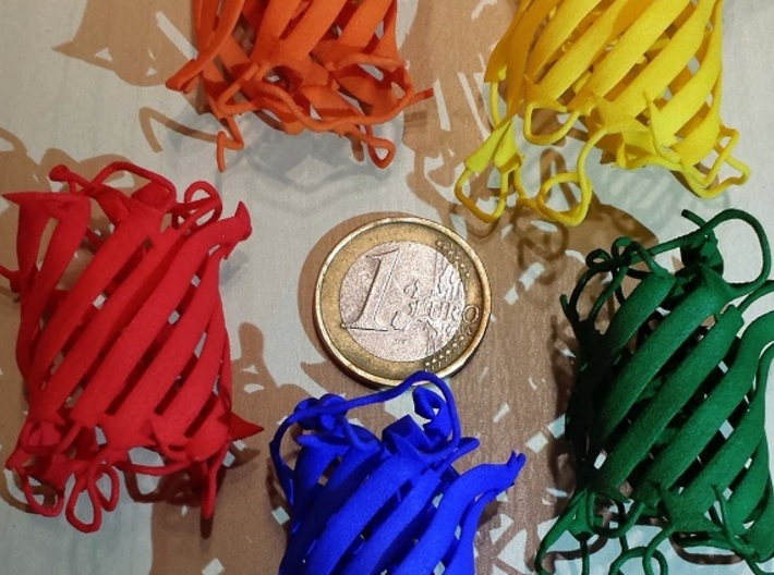
I picked five fluorescent proteins to represent the wide array that have been engineered over the years. The original GFP from jellyfish [9] was mutated to make the blue Cerulean and the yellow Citrine [10,11]. The coral protein RFP was then
converted into mCherry and mOrange, which are both part of the larger collection of "mFruits" from the Tsien lab [12]. Each model shows the barrel structure that protects the chromophore running through the center. Get your hands on the models to appreciate the subtle differences that make each protein unique! Each model comes in its respective color; I've also offered each model in White Strong & Flexible in case you would like to hand paint it. Just make sure you don't mix them up before they're painted!
Sources for the curious:
[1]* Osamu Shimomura, "Discovery of Green Fluorescent Protein, GFP." Nobel Lecture (2008)
[2]* Martin Chalfie, "GFP: Lighting Up Life." Nobel Lecture (2008)
[3]* Roger Tsien, "Constructing and Exploiting the Fluorescent Protein Paintbox." Nobel Lecture (2008)
[4] W.D. McElroy and Arda Green, "Function of adenosine triphosphate in the activation of luciferin." Archives of Biochemistry and Biophysics (1956)
[5]* Osamu Shimomura, "Structure of the chromophore of aequorea green fluorescent protein." FEBS Letters (1979)
[6] Martin Chalfie et al., "Green fluorescent protein as a marker for gene expression." Science (1994)
[7]* Roger Heim and Roger Tsien, "Engineering green fluorescent protein for improved brightness, longer wavelengths and fluorescence resonance energy transfer." Current Biology (1996)
[8]* Robert Campbell et al, "A monomeric red fluorescent protein." PNAS (2002)
[9] Mats Ormö et al, "Crystal structure of the Aequorea victoria green fluorescent protein." Science (1996)
[10] Mickaël Lelimousin et al, "Intrinsic Dynamis in ECFP and Cerulean Control Fluorescence Quantum Yield." Biochemistry (2009)
[11]* Oliver Griesbeck et al, "Reducing the Environmental Sensitivity of Yellow Fluorescent Protein." Journal of Biological Chemistry (2001)
[12] Xiaokun Shu et al, "Novel Chromophores and Buried Charges Control Color in mFruits." Biochemistry (2006)
Details


More From This Shop